- Application
- Lid margin and eyelash imaging for Demodex blepharitis
- Clinical Domain
- Anterior segment and ocular surface, including blepharitis assessment and treatment monitoring
- Relevant For
- Device makers, Solution builders, Researchers, Pharma and biotech, and CROs
- What Teams Can Do
- Detect, Track, and Study
- Platform Layers
- Ocunur headset, eye imaging, controlled illumination, SDK, middleware privacy controls, and IntelliMDM
Overview
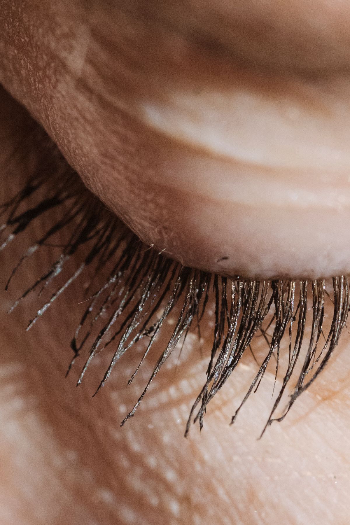
Demodex blepharitis is an inflammation of the eyelid margin caused by an overpopulation of Demodex mites at the base of the eyelashes. It is one of the most common causes of blepharitis and is often missed during routine exams. The defining clinical sign is collarettes, which are cylindrical deposits that form around the base of the lashes. With a prescription treatment approved by the FDA in 2023, the condition now has a clear path from identification to therapy, which increases the value of consistent, repeatable lid margin imaging.
This use case describes how teams can use the IntelliXR® platform to capture standardized images of the lid margin and eyelashes, store them securely, compare them over time, and run the same capture protocol across many sites.
Clinical Context
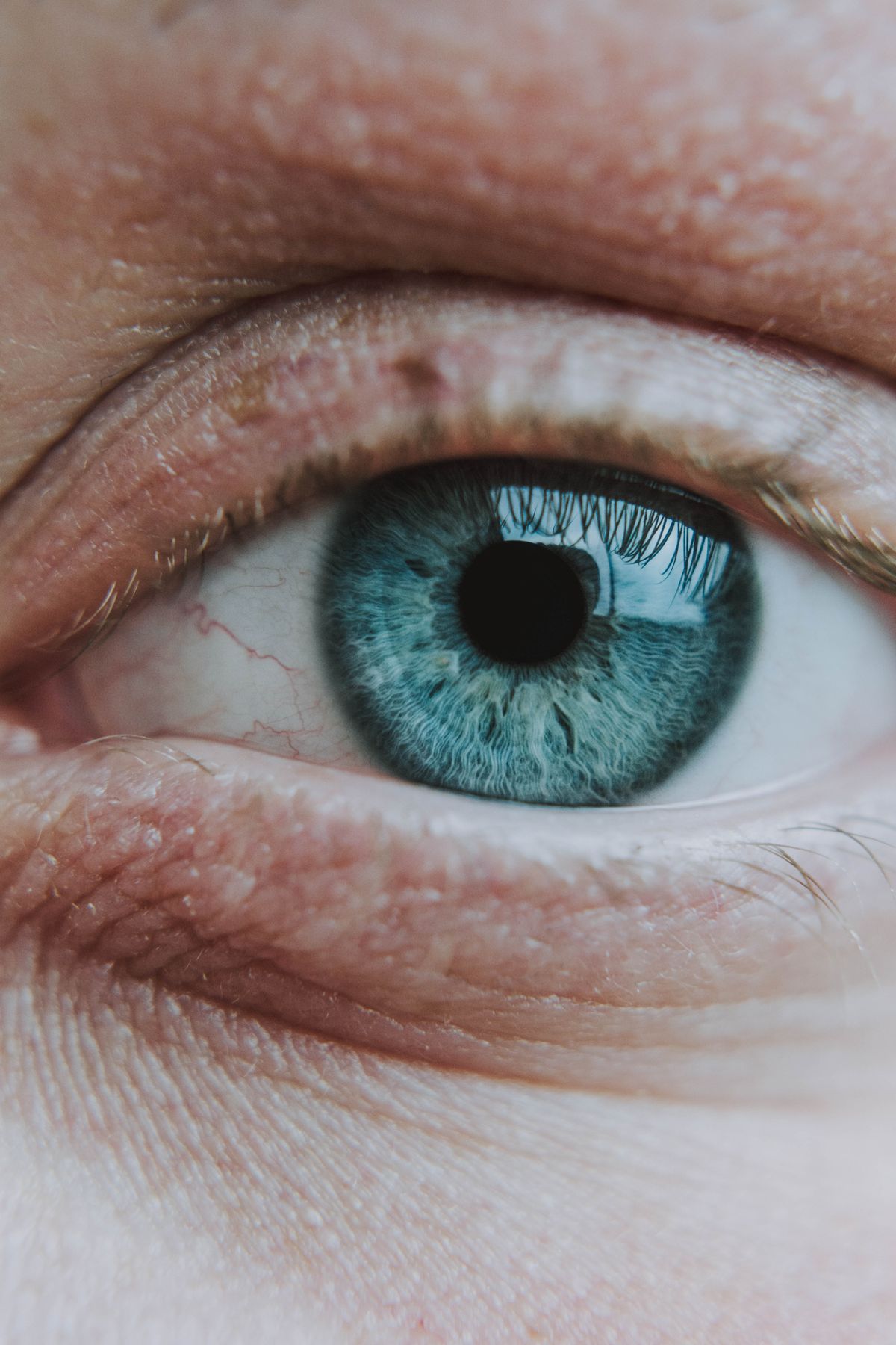
Today, Demodex blepharitis is assessed at the slit lamp. A clinician asks the patient to look down, inspects the base of the upper lashes for collarettes, and grades what they see. Confirmation can involve removing lashes and counting mites under a microscope.
This approach depends on the equipment available in the room and on the individual examiner. Lighting, magnification, and patient position vary between visits, which makes it harder to compare results over time or across sites.
A head-mounted approach gives teams a consistent capture environment. The headset fixes the distance between camera and eye, controls the illumination, and runs the same protocol for every patient.
What Teams Can Do
1. Detect
Capture lid margin and lash images during a standard eye exam or screening session, and flag collarettes for clinician review.
2. Track
Repeat the same capture protocol at each visit to compare lid margin images before, during, and after treatment.
3. Study
Collect standardized, de-identified image sets across sites for clinical trials, observational studies, and algorithm development.
4. Combine
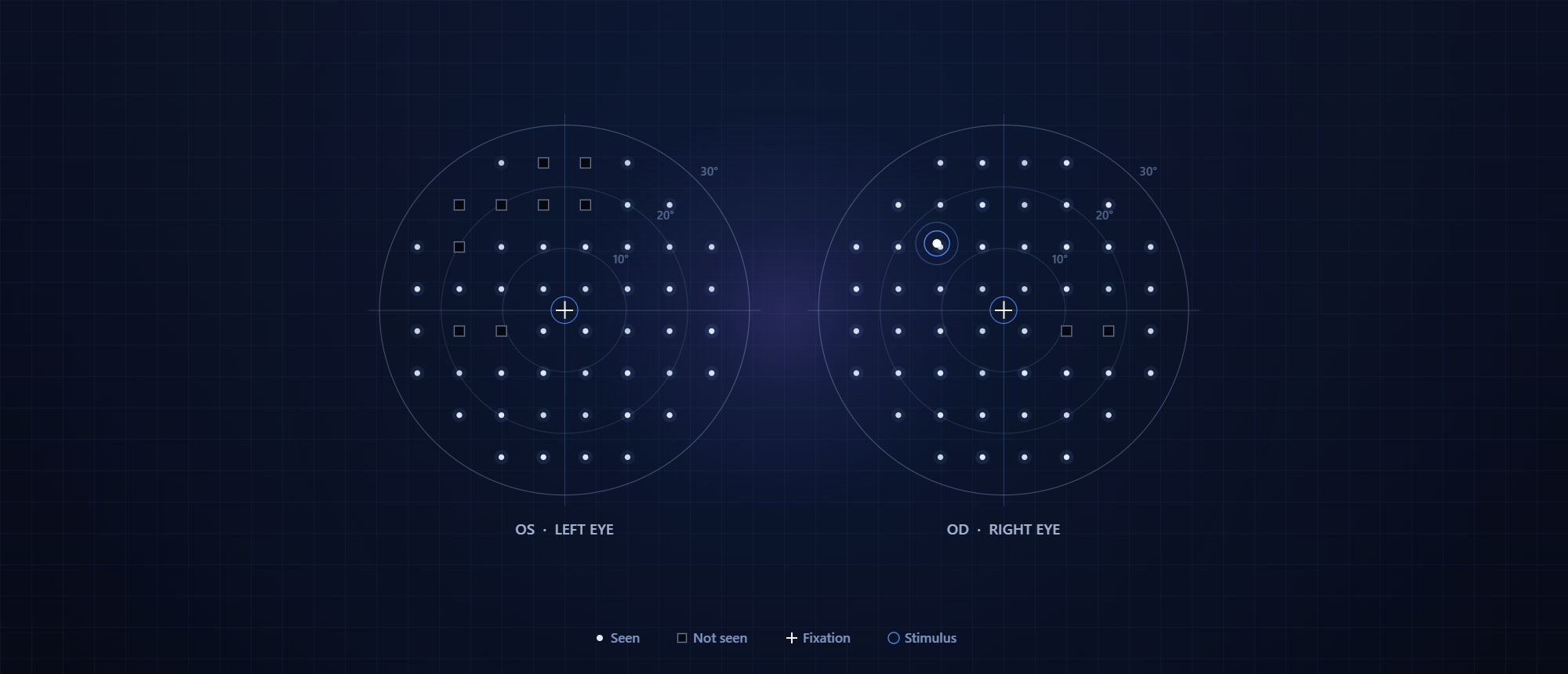
Run lid margin imaging in the same session as other platform applications, such as dry eye assessment or visual field testing, on the same headset.
Requirements and Platform Capabilities
| Requirement | Why It Matters | Platform Capability |
|---|---|---|
| Consistent image geometry | Images must be comparable across visits and patients | Fixed camera-to-eye distance inside the Ocunur headset |
| Controlled illumination | Collarettes are judged by appearance, which changes with lighting | Illumination controlled by the application through the SDK |
| Guided patient positioning | The base of the upper lashes is visible only when the patient looks down | On-display fixation targets that direct gaze during capture |
| Repeatable protocols | Treatment tracking and trials need the same capture every time | Capture protocols defined in the application and applied on every device |
| Structured session records | Images must link to study, subject, and visit | Sessions bound to anonymized study and subject identifiers, exported as JSON or CSV |
| Patient data protection | Eye images are sensitive clinical data | De-identification, metadata stripping, AES-256 encryption, consent, and audit logging |
| Multi-site consistency | Trials and clinic networks need the same configuration at every site | IntelliMDM enrollment, policy enforcement, and OTA updates |
System Architecture
A Demodex blepharitis application sits at Tier 4 of the IntelliXR® stack. Every layer beneath it is provided by the platform.
| Tier | Layer | Role in Lid Margin Imaging |
|---|---|---|
| Tier 4 | Client Application | Capture protocol, image review, grading workflow, and results display. |
| Tier 3 | SDK | Eye imaging, display, session, data, privacy, and device modules. |
| Tier 2 | Middleware Runtime | Image handling, encryption, de-identification, and session management. |
| Tier 1 | Hardware Abstraction Layer | Eye camera, illumination, and display drivers. |
| Tier 0 | Hardware | Ocunur headset and Android compute host. |
| Fleet | IntelliMDM | Device enrollment, policy enforcement, security, and OTA updates across sites. |
How It Works
6.1 Session Setup
Each capture runs as a session bound to anonymized study and subject identifiers. The application loads its capture protocol, which defines illumination settings, gaze targets, and the number of images per eye.
6.2 Guided Gaze
The headset display presents a fixation target that moves the patient’s gaze downward, which exposes the base of the upper lashes to the camera. The same targets are used at every visit, so the lid margin is captured from a consistent angle.
6.3 Image Capture
The eye imaging module captures images of the lid margin and lashes under the illumination set by the application. Each image is timestamped and linked to the session, the eye, and the gaze target in use at the moment of capture.
6.4 Review and Grading
Images are available to the application for clinician review and grading. Teams developing automated analysis can use the same images as inputs to their own detection or grading models.
6.5 Longitudinal Comparison
Because geometry, lighting, and gaze are held constant, images from different visits can be placed side by side to follow changes in collarettes and lid margin appearance through treatment.
6.6 Data Handling
Privacy controls run on the device. Eye images are de-identified, metadata is stripped, exports are encrypted with AES-256, and every access is recorded in a tamper-evident audit log before data leaves the headset.
6.7 Fleet Management
Every headset is enrolled in IntelliMDM, which applies the same capture configuration and security policies at every site. Protocol updates reach the full fleet remotely, so every site moves to the same version together.
Example Applications
1. Screening in routine eye exams
A solution builder adds lid margin imaging to an existing headset-based exam, so collarettes can be flagged during the same visit as other tests.
2. Treatment response monitoring
A clinic tracks patients on Demodex treatment by repeating the same capture at each follow-up visit and comparing images over time.
3. Clinical trial endpoints
A pharma sponsor or CRO uses standardized lid margin images as a consistent endpoint across trial sites, with de-identified data collected centrally.
4. Algorithm development
A research team builds a labeled image set for training and validating automated collarette detection and grading models.
Key Takeaways
1. Standardized capture makes lid margin images comparable
Fixed geometry, controlled illumination, and guided gaze give every image the same conditions, visit after visit.
2. One protocol supports detection, tracking, and research
The same capture workflow serves screening, treatment monitoring, and clinical studies.
3. Privacy is handled at the point of capture
De-identification and encryption run on the device, so sensitive eye images are protected before they leave the headset.
4. Fleet management keeps every site aligned
IntelliMDM keeps capture protocols and security policies consistent across clinics and trial sites.
5. The same headset supports other ocular surface applications
Lid margin imaging runs on the same platform as dry eye assessment and other vision diagnostics.
Develop ocular surface applications on the IntelliXR® platform. Book a demo or explore the IntelliXR® technical documentation.